
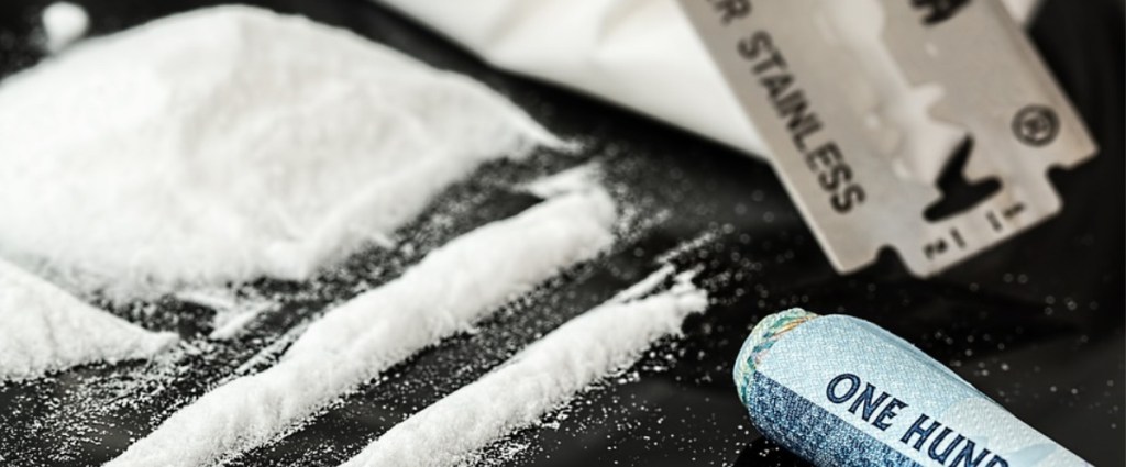

The fact that mind-altering chemicals like cocaine do a number on our bodies isn’t exactly breaking news, but these MRI’s show just how far cocaine can take a person beyond pounding hearts, sweaty palms, and strained nerves.
In fact, they tell us that repeated and heavy abuse of the drug can lead to physical brain changes – the white matter being eaten away – that can be permanent.
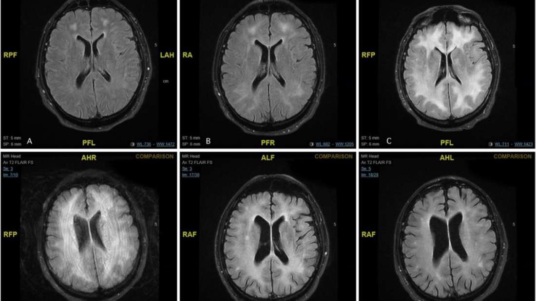
Image Credit: BMJ Case Reports
The scans come from a 45-year0old man who arrived in a Maltese emergency room reporting confusion and unusual behavior. The doctor who treated the man, Dr. Ylenia Abdilla, said in the study…
“The patient was not cooperative, unable to perform simple tasks and was not following commands. He was moving all four limbs in purposeful movement.”
Things took a turn for the worse when he stopped being able to communicate and then became catatonic, and even though his blood tests and exams returned normal, his MRI scan was definitely a source of concern.
Doctor’s diagnosed him with leucoencephalopathy, the progressive damage or inflammation of the white matter of the brain. The condition is rare, and typically associated with infection – but the man didn’t have one.
https://www.instagram.com/p/BDwvBQ8n9-d/
Even though the man claimed to not have used cocaine for 2 to 3 days, he was a regular user and significant quantities showed up in his urine, leading doctors to assume the condition was a result of the abuse. Doctors treated him with drugs used to decrease inflammation, boost his immune system, and remove antibodies from blood plasma.
Similar cases of cocaine abuse leading to leucoencephalopathy have been documented a handful of times, though doctors and researchers aren’t clear on why cocaine has such an effect on the brain. Some studies suggest that heavy drug use can result in a “splitting and unraveling” of the layer that’s supposed to insulate nerves and reduce swelling.
So what happened to the man in the study? Though often fatal, the man in this case not only recovered, but scored normally on neurological tests a year later. His scans reveal that the white matter has not regenerated or repaired itself, however, even though he has completed a drug rehab program.
“He had not used drugs for 1 year. Apart from some complaints of low mood, he was fully independent and had returned to his previous functional status,” the study concluded.


